CAMERA TEMPLATE
Ideal solution for a review board, teaching setting, collaborative environments, and industrial applications.
The HD-210U is an innovative HD brightfield microscopy camera designed for maximum flexibility and ease of use. The HD-210U offers full, high definition, 1080p60 HDMI streaming with superb color reproduction as well as USB2 connectivity for simultaneous live viewing and image capture.
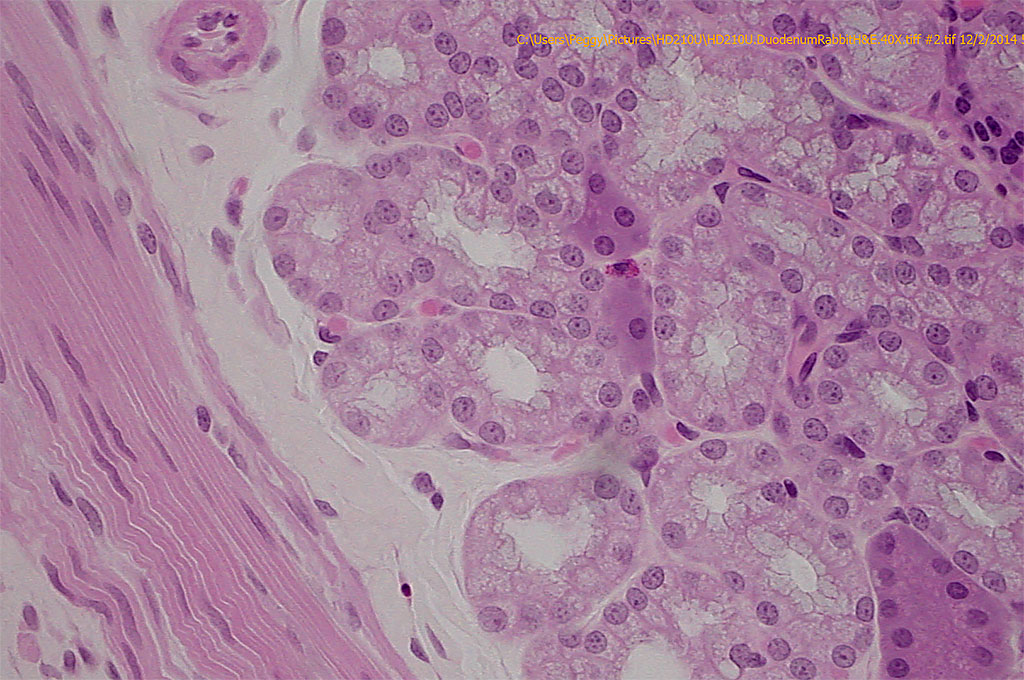
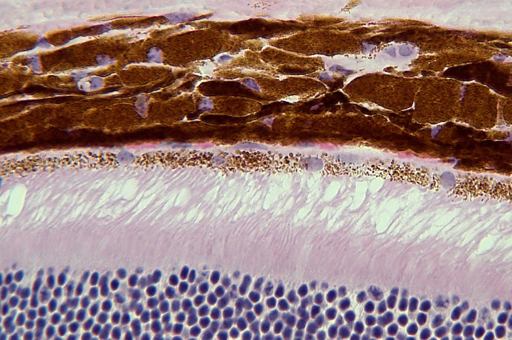
The HD-210U is preprogrammed for perfect brightfield color by critical adjustments of 6 hues – not just red, green and blue, but yellow, cyan and magenta as well. Real time, fast 60fps in live streaming mode enables the operator to move samples or change magnifications without smear lag or jitter. Real-time, fast 60fps video streaming allows effortless focus and specimen placement for more efficient slide reviews.
The HD-210U automatically adjusts to changes in light intensity. No more fumbling with the intensity controls of your microscope or camera. One button white balance and preprogrammed scene files makes using the HD-210U a snap to use. Operate the camera by directly connecting to an HDMI monitor without the need for a PC, or utilize the USB 2.0 output for image capture.
Free MagicApp acquisition software is included with the HD-210U.
The Dage-MTI HD-210U is a complete, cost-effective, high quality solution for life science, clinical science, educational or industrial microscopy applications.
- Cost-effective, high quality solution.
- Color bar test signal – allows the user to quickly and correctly set the contrast and brightness on a monitor
- Exceptional color reproduction. Color calibration utilizing Cyan, Magenta, Yellow, Red, Green, Blue (CMYRGB) for 6 color accuracy vs RGB.
- 1080p60 high definition video streaming over HDMI connector for live viewing without the need for a computer.
- Simultaneous USB2 output for image capture, if desired.
- Easy to use – 1 button white balance. Factory preprogrammed menu settings for optimal results.
- High resolution color 2.1 megapixel camera with 1/3 format CMOS sensor.
- MagicApp software (included) set up in minutes for image capture (Win XP (SP2-3)/Vista/7/8).
- One click image capture and instantaneous review.
- Image capture in TIF, JPEG, Bitmap, PNG.
- One (1) year parts and labor warranty.
- Pathology
- Education
- Tumor Boards
- Clinical and Industrial Microscopy
- Brightfield
- Live Cell Imaging/Classroom demonstrations
- Live dissections or animal surgeries
- Lab Practicals
- Histology
- Cytology
- Defect Analysis
- Semiconductor Inspection
- Metrology
| Specification | |
| Image sensor | 1/3 inch color CMOS sensor, 2.1 megapixel |
| Output pixels | Horizontal: 1920, Vertical: 1080 |
| Signal system | 1080/59.94p, 1080/50p, 1080/59.94i, 1080/50i, 720/59.94p, 720/50p |
| Sensitivity | F 4 standard (2000 lx, 3000 K) |
| Minimum illumination | 8 lx standard (50 IRE, F1.4 gain + 18 dB, gamma setting ON (setting value 0), 3000 K) |
| Output signal | DVI (Digital RGB) DVI-D terminal USB Video Class 1.1 mini-USB terminal (in conformity witd USB 2.0) |
| Sync system | Internal |
| White balance | ATW (Automatic tracking white balance), AWB (Automatic white balance), MANUAL (Manual) |
| Gain | MANUAL (Manual), OFF (0 dB) |
| Scene file |
|
| Lens mount | C mount |
| Power supply | 12V DC+10% |
| Power consumption | Approx 4.1 w |
| Weight | Approx 148g (0.326 lbs) |
| External dimension | 44(W) x 44(H) x 78(D) mm (1.73”(W) x 1.73”(H) x 3.07”(D)) |
| Operating temperature | 0 °C to 40 °C (32° F to 104° F) |
| Operating humidity | Less tdan 90% (non condensing) |
*Design and specifications are subject to change without notice
OS: Windows® XP SP2 – SP3, Windows® Vista, Windows® 7
CPU: Intel® Pentium® compatible (x86 or x64) CPU, 1 GHz or more
Memory: 512 MB or more
USB Port: USB 2.0


