Electrophysiology & Deep Tissue Imaging
Enhanced Capability for Electrophysiology and Deep Tissue Imaging
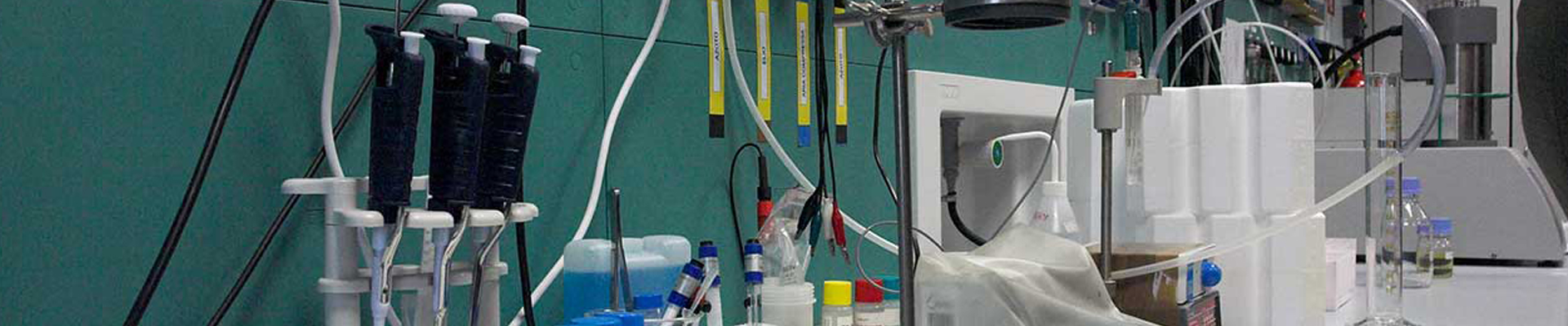
Electrophysiology can entail unique imaging challenges.
- Dim, low-contrast imaging conditions due to light scatter within the tissue.
- Little to no visible light in deeper tissue imaging environments.
- Fast, real time frame rates (30fps) required for accurate placement of micropipettes/electrodes.
Meeting the Challenge
The IR-1000 is a real-time camera solution that offers enhanced sensitivity across the entire visible and near IR spectrum. The IR-1000 boasts a 5X increase in sensitivity at 900nm – all at 30 fps. Its sensitivity is ideal for visible, IR, near IR and bright fluorescence modes of operation. The wavelength used by the operator determines tissue observation.
- Visible Light DIC/Dodt gradient contrast imaging – Allows operator high resolution observation of the tissue surface.
- 775nm IR-DIC/Dodt gradient contrast imaging – Allows observation within the tissue slice
- 900nm IR- DIC/Dodt gradient contrast imaging – Allows observation deeper into the tissue.
Important features of the IR-1000 camera include: Automatic Contrast and real-time edge enhancement. When a scene changes, the camera’s electronics automatically and instantaneously readjust to achieve optimum contrast. There is no need for recalibration by the operator. The enhancement function provides an edge sharpening of soft IR images, resulting in a clearer image.
For increased sensitivity, on-chip gating can be accomplished using a qualified frame grabber board or the Investigater from Dage-MTI – a stand-alone controller providing real-time gating without the need for a computer.
Related Products
[DAGE_RELATED_PRODUCTS solutions=’electrophysiology-and-deep-tissue’]